Skip Navigation
Skip to contents

JYMS : Journal of Yeungnam Medical Science

Indexed in: ESCI, Scopus, PubMed,
PubMed Central, CAS, DOAJ, KCI
FREE article processing charge
OPEN ACCESS
SEARCH
Search

Articles

Page Path
HOME > J Yeungnam Med Sci > Volume 40(2); 2023 > Article
Resident fellow section: Teaching images
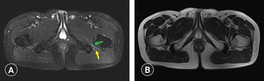
A 40-year-old man with neuropathic pain in the entire left foot
Jae Hwa Bae1,*orcid, Mathieu Boudier-Revéret2orcid, Min Cheol Chang1orcid
Journal of Yeungnam Medical Science 2023;40(2):223-224.
DOI: https://doi.org/10.12701/jyms.2022.00486
Published online: August 30, 2022

1Department of Physical Medicine and Rehabilitation, Yeungnam University College of Medicine, Daegu, Korea

2Department of Physical Medicine and Rehabilitation, Centre Hospitalier de l’Université de Montréal, Montreal, QC, Canada

Corresponding author: Min Cheol Chang, MD Department of Physical Medicine and Rehabilitation, Yeungnam University College of Medicine, 170 Hyeonchung-ro, Nam-gu, Daegu 42415, Korea Tel: +82-53-620-4682 • Fax: +0504-231-8694 • E-mail: wheel633@gmail.com
*The first author, Jae Hwa Bae, is currently in training.
• Received: July 13, 2022   • Revised: July 24, 2022   • Accepted: July 31, 2022

Copyright © 2023 Yeungnam University College of Medicine, Yeungnam University Institute of Medical Science

This is an Open Access article distributed under the terms of the Creative Commons Attribution Non-Commercial License (http://creativecommons.org/licenses/by-nc/4.0/) which permits unrestricted non-commercial use, distribution, and reproduction in any medium, provided the original work is properly cited.

  • 1,241 Views
  • 59 Download
A 40-year-old man with no medical history visited our clinic because of pain in the entire left foot that had persisted for 1 year. The pain was aggravated by sitting, walking, running, and climbing stairs. The pain was stabbing, burning, and tingling and was rated at 7 on a numeric rating scale. In addition to left foot pain, he reported mild left buttock pain with no history of left lower limb or pelvic injury. Physical examination revealed no motor or sensory deficits, and deep tendon reflexes were normal in all four limbs. His straight leg raise test results were negative. Tinel sign over the left tibial nerve in the tarsal tunnel was negative and his foot pain was not aggravated by ankle inversion/eversion.
Differential diagnoses included lumbosacral radicular pain due to spinal disorders, tarsal tunnel syndrome, and sciatic neuropathy. Lumbar magnetic resonance imaging (MRI) showed a diffuse bulging disc on L4–L5 and L5–S1 with a high-intensity zone. No pain reduction was observed on diagnostic blocks of the left L5 and S1 nerve roots and left tibial nerve within the tarsal tunnel with 1 mL of 2% lidocaine. A nerve conduction study and electromyography of the left lower limb revealed no abnormalities.
Further investigation was conducted. On pelvic axial fat-saturated T2-weighted MRI, high signal intensity was found in the left quadratus femoris muscle (Fig. 1). Additionally, the ischiofemoral space was narrower on the left than on the right side. The sciatic nerve was located over an area with high signal intensity. The patient was diagnosed with left ischiofemoral impingement. For the injection procedure, the patient was placed in a prone position, and the left buttock was scanned with a 6-MHz curved probe (LOGIQ P6, General Electric, Seoul, Korea) to acquire axial images. A mixed solution of 20-mg triamcinolone, 1-mL 1% lidocaine, and 3.5-mL normal saline was administered via ultrasound-guided injection to the left sciatic nerve and quadratus femoris muscle. The patient’s pain was alleviated by 80% at the 3-week follow-up examination.
Ischiofemoral impingement is an uncommon cause of buttock pain due to impingement of the quadratus femoris muscle between the lesser trochanter and lateral border of the ischium [1-3]. Most patients with ischiofemoral impingement experience buttock pain [1-3]. Neuropathic pain radiating towards the posterior aspect, calf, and foot can occur due to irritation of the adjacent sciatic nerve. There are only a few reports on the diagnosis and treatment of this condition. When conservative treatments with activity modification, stretching of the hip muscles, and ischiofemoral space injection are ineffective, surgical treatment (resection of the lesser trochanter) can be considered [1-3].
In the present case, pain throughout the left foot was caused by ischiofemoral impingement. Clinicians should consider this possibility in patients with neuropathic pain radiating to the area innervated by the sciatic nerve, combined with buttock pain and aggravated by hip motion.
• Ischiofemoral impingement can cause buttock pain with or without sciatica due to irritation of the adjacent sciatic nerve, which is caused by impingement of the quadratus femoris muscle between the lesser trochanter and lateral border of the ischium.
• Edema and a narrowed ischiofemoral space identified by pelvic MRI indicate ischiofemoral impingement.
• For ischiofemoral impingement, conservative treatment with activity modification and ischiofemoral space injection can be attempted. In cases that are refractory to conservative treatment, surgical treatment should be considered.

Ethical statements

This study was approved by the Institutional Review Board (IRB) of Yeungnam University Hospital (IRB No: 2022-07-029). Written informed consent was obtained for the publication of this report.

Conflicts of interest

Mathieu Boudier-Revéret has been editorial board member of Journal of Yeungnam Medical Science (JYMS) since 2021. Min Cheol Chang has been Associate editor of JYMS since 2021. They were not involved in the review process of this manuscript. Otherwise, there is no conflict of interest to declare.

Funding

None.

Author contributions

Conceptualization: MBR, MCC; Data curation, Formal analysis, Methodology, Visualization, Investigation, Resources, Supervision, Validation: MCC; Writing-original draft: all authors; Writing-review & editing: all authors.

Fig. 1.
(A) Pelvic axial fat-saturated T2-weighted and (B) T2-weighted magnetic resonance imaging (MRI) reveal that the ischiofemoral space is narrower on the left side than on the right side. Additionally, on the pelvic axial fat-saturated T2-weighted MRI (A), high signal intensity is found on the left quadratus femoris muscle (green arrow), indicative of muscle edema. Over the area of high signal intensity in the left quadratus femoris muscle, the left sciatic nerve is visible (yellow arrow).
jyms-2022-00486f1.jpg
  • 1. Lee S, Kim I, Lee SM, Lee J. Ischiofemoral impingement syndrome. Ann Rehabil Med 2013;37:143–6.ArticlePubMedPMC
  • 2. Ulusoy OL, Tutar S, Ozturk E, Mutlu A, Mutlu H. Ischiofemoral impingement syndrome: another cause of extraspinal sciatica. Spine J 2016;16:e527.ArticlePubMed
  • 3. Wilson MD, Keene JS. Treatment of ischiofemoral impingement: results of diagnostic injections and arthroscopic resection of the lesser trochanter. J Hip Preserv Surg 2016;3:146–53.ArticlePubMedPMC

Figure & Data

References

    Citations

    Citations to this article as recorded by  

      Figure

      JYMS : Journal of Yeungnam Medical Science