E-Submission
PubMed Central, CAS, DOAJ, KCI

Articles
- Page Path
- HOME > J Yeungnam Med Sci > Volume 37(3); 2020 > Article
-
Case report
Spontaneous resolution of serous retinal detachment caused by choroidal mass after a first trimester abortion -
You Hyun Lee
 , Yu Cheol Kim
 -
Yeungnam University Journal of Medicine 2020;37(3):242-245.
DOI: https://doi.org/10.12701/yujm.2020.00087
Published online: April 6, 2020
Department of Ophthalmology, Keimyung University Dongsan Hospital, Keimyung University School of Medicine, Daegu, Korea
- Corresponding author: Yu Cheol Kim Department of Ophthalmology, Keimyung University Dongsan Hospital, Keimyung University School of Medicine, 1035 Dalgubeoldae-ro, Dalseo-gu, Daegu 42601, Korea Tel: +82-53-258-4545 Fax: +82-53-258-7130 E-mail: eyedr@dsmc.or.kr
Copyright © 2020 Yeungnam University College of Medicine
This is an Open Access article distributed under the terms of the Creative Commons Attribution Non-Commercial License (http://creativecommons.org/licenses/by-nc/4.0/) which permits unrestricted non-commercial use, distribution, and reproduction in any medium, provided the original work is properly cited.
- 5,098 Views
- 65 Download
- 2 Crossref
Abstract
- Pregnancy-related ocular diseases develop mostly in the third trimester of pregnancy. Here, we describe a case of a pregnant woman with a choroidal mass that caused a serous retinal detachment during the first trimester of pregnancy. The patient’s condition resolved spontaneously after an abortion.
- During pregnancy, cardiac output increases and hormone levels fluctuate. These physiological changes in pregnant women often cause retinal and choroidal diseases or worsen preexisting retinal and choroidal conditions [1,2]. For example, the incidence of central serous chorioretinopathy (CSC) and circumscribed choroidal hemangioma (CCH) increase during pregnancy [3,4]. Specifically, these diseases become exacerbated during the third trimester and spontaneously regress after childbirth [5,6]. Here, we present a case of a pregnant woman with a choroidal mass causing serous retinal detachment (SRD) during the first trimester of pregnancy that resolved spontaneously after an abortion.
Introduction
- This study was approved by the Institutional Review Board of the Keimyung University Dongsan Hospital (IRB No: DSMC 2020-02-070). The patient provided written informed consent for publication of clinical details and images.
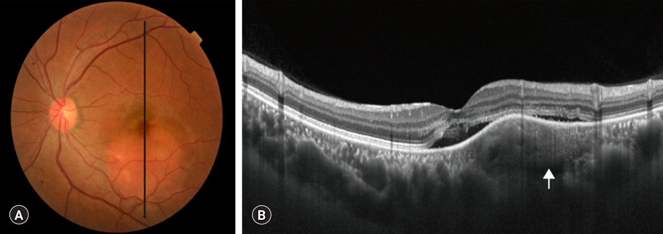
- A 40-year-old woman was referred to our hospital with a 3-day history of visual disturbance in her left eye. The patient was a primigravida who was in her first trimester of pregnancy (7 weeks) at the time of referral. She did not have gestational hypertension or diabetes. Her left eye initially had a best-corrected visual acuity (BCVA) of 20/50 (according to the Snellen chart) and an intraocular pressure (IOP) of 15 mmHg. Her right eye had a BCVA of 20/20 and an IOP of 17 mmHg. Examination of the fundus revealed an orange-red retina in the left eye with posterior pole elevation (Fig. 1A).
- Optical coherence tomography (OCT; DRI OCT Triton, Topcon, Tokyo, Japan) revealed a thick (>500 µm) elevated choroid, an SRD, compression of the choriocapillaris, and a choroidal mass located between the choriocapillaris and the outer choroidal tissue in the left eye (Fig. 1B). We avoided fluorescein angiography (FA) and indocyanine green angiography (ICGA) because of their possible teratogenicity. The patient was diagnosed with a choroidal mass. The appearance of the mass suggested possible CCH. We decided to closely monitor the changes in the patient’s BCVA and the progression of her SRD.
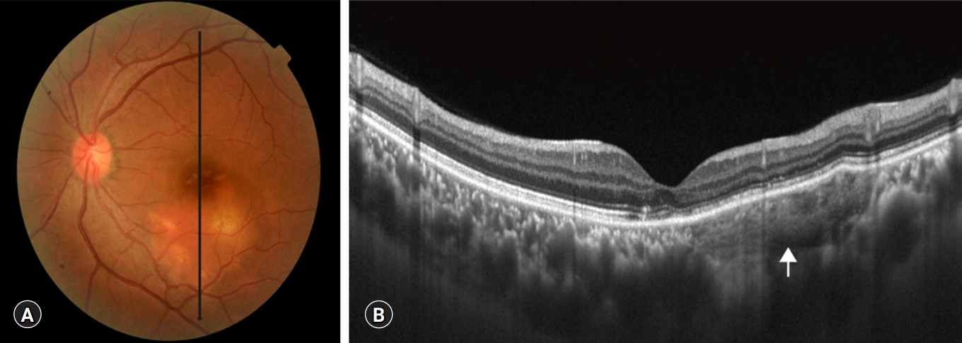
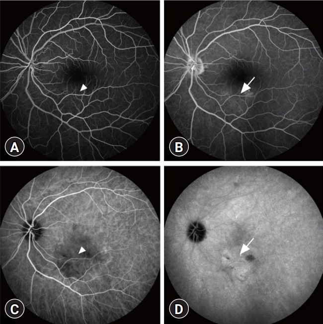
- One month later, the patient revisited our clinic after having had an abortion during her 9th week of pregnancy. Her left-eye BCVA was 20/20 and her IOP was 14 mmHg. Examination of the fundus showed that the elevated retinal height had decreased and that the retinal surface exhibited a patchy yellowish discoloration (Fig. 2A). OCT revealed a decreased SRD with visible fine lamellar lines of the choroidal lesion (Fig. 2B). FA (HRA-2; Heidelberg Engineering, Dossenheim, Germany) revealed patchy hyperfluorescence in the area corresponding with the previously elevated retina during the arteriovenous phase (Fig. 3A) with late-phase staining (Fig. 3B). ICGA revealed early hypofluorescence (Fig. 3C), followed by diffuse confluent fluorescence during the late phase (Fig. 3D). We requested that the patient visit the hospital 3 months later, however she was lost during this follow-up period.
Case
- CCH appears as an indistinct, round-to-oval, orange-red mass located posterior to the equator of the eye [6]. CSC is characterized as an accumulation of subretinal fluid that causes circumscribed neurosensory retinal detachment [7]. Distinguishing between these diseases during pregnancy is difficult because using either FA or ICGA is discouraged during this period owing to their possible teratogenicity [5]. In this case, at 7 weeks of pregnancy, we initially suspected CCH because of the lesion’s orange-red elevated appearance, the presence of SRD, and the presence of a thick and elevated choroid with an associated mass. Additionally, we eliminated the possibility of CSC as it typically develops in women with pre-eclampsia [8]. FA and ICGA were performed after the patient’s abortion. FA revealed early patchy hyperfluorescence with late-phase staining, and ICGA revealed early hypofluorescence with later diffuse confluent fluorescence. The FA findings are consistent with the CCH diagnosis, as CCH is characterized by a stippled choroidal hyperfluorescence followed by increasing hyperfluorescence and progressive staining of the tumor. However, these FA characteristics are not pathognomonic for CCH. Unlike the FA findings, the ICGA findings are inconsistent with the CCH diagnosis, as CCH manifests as extreme early hyperfluorescence that decreases in the later phases (known as the "washout" phenomenon) [9]. Compression of the choriocapillaris was visible upon the initial OCT, however CCH typically develops in the absence of choriocapillaris compression [10].
- Choroidal osteomas are benign, rare, ossifying choroidal tumors that typically affect the juxtapapillary and macular areas of young healthy females [11]. The color of the choroidal osteoma depends on the depigmentation of the retinal pigment epithelium [13]. In our case, an orange-red lesion in the fundus was seen at an early stage, which is in line with the diagnostic features of CCH. The fine lamellar lines found within the choroidal mass during OCT after the patient’s abortion indicated the possible presence of an early choroidal osteoma. Moreover, ICGA of choroidal osteoma usually reveals early hypofluorescence and late-phase hyperfluorescence [12]. However, we did not perform B-scan ultrasonography in this case, which was consistent with a previous study [14]. After the patient’s abortion her decreased visual acuity improved and her SRD resolved, which implies that her pregnancy was directly related to the growth of the choroidal mass. While the mechanism remains unclear, we postulated that the increase in maternal cardiac output during the first trimester may have caused hyperpermeability of the vascular channels within the choroidal mass, which may have led to its exaggerated appearance [15].
- In conclusion, it is possible for SRD to develop in first trimester of pregnancy along with an associated choroidal mass. This SRD will most likely resolve spontaneously after an abortion or the termination of pregnancy. Our finding highlights the direct correlation between pregnancy and choroidal masses, which may help physicians and specialists anticipate these issues in pregnant patients.
Discussion
-
Conflicts of interest
No potential conflict of interest relevant to this article was reported.
-
Funding
This work was supported by the National Research Foundation of Korea grant (NRF-2019R1G1A1011559).
-
Author contributions
Conceptualization: YHL, YCK; Data curation: YHL; Formal analysis: YCK; Supervision: YCK; Writing-original draft: YHL; Writing-review & editing: YCK.
Notes

- 1. Thornburg KL, Jacobson SL, Giraud GD, Morton MJ. Hemodynamic changes in pregnancy. Semin Perinatol 2000;24:11–4.ArticlePubMed
- 2. Errera MH, Kohly RP, da Cruz L. Pregnancy-associated retinal diseases and their management. Surv Ophthalmol 2013;58:127–42.ArticlePubMed
- 3. Chatziralli I, Kabanarou SA, Parikakis E, Chatzirallis A, Xirou T, Mitropoulos P. Risk factors for central serous chorioretinopathy: multivariate approach in a case-control study. Curr Eye Res 2017;42:1069–73.ArticlePubMed
- 4. Cohen VM, Rundle PA, Rennie IG. Choroidal hemangiomas with exudative retinal detachments during pregnancy. Arch Ophthalmol 2002;120:862–4.PubMed
- 5. Sayman Muslubas I, Arf S, Hocaoglu M, Ozdemir H, Karacorlu M. Spontaneous regression of serous retinal detachment associated with circumscribed choroidal hemangioma after childbirth. Retin Cases Brief Rep 2017;11:7–11.ArticlePubMed
- 6. Heimann H, Damato B. Congenital vascular malformations of the retina and choroid. Eye (Lond) 2010;24:459–67.ArticlePubMedPDF
- 7. Daruich A, Matet A, Dirani A, Bousquet E, Zhao M, Farman N, et al. Central serous chorioretinopathy: recent findings and new physiopathology hypothesis. Prog Retin Eye Res 2015;48:82–118.ArticlePubMed
- 8. Morikawa M, Cho K, Kojima T, Chiba K, Ishikawa S, Umazume T, et al. Risk factors for central serous chorioretinopathy in pregnant Japanese women. J Obstet Gynaecol Res 2017;43:866–72.ArticlePubMed
- 9. Arevalo JF, Shields CL, Shields JA, Hykin PG, De Potter P. Circumscribed choroidal hemangioma: characteristic features with indocyanine green videoangiography. Ophthalmology 2000;107:344–50.ArticlePubMed
- 10. Rojanaporn D, Kaliki S, Ferenczy SR, Shields CL. Enhanced depth imaging optical coherence tomography of circumscribed choroidal hemangioma in 10 consecutive cases. Middle East Afr J Ophthalmol 2015;22:192–7.ArticlePubMedPMC
- 11. Shields CL, Shields JA, Augsburger JJ. Choroidal osteoma. Surv Ophthalmol 1988;33:17–27.ArticlePubMed
- 12. Gass JD, Guerry RK, Jack RL, Harris G. Choroidal osteoma. Arch Ophthalmol 1978;96:428–35.ArticlePubMed
- 13. Lafaut BA, Mestdagh C, Kohno T, Gaudric A, De Laey JJ. Indocyanine green angiography in choroidal osteoma. Graefes Arch Clin Exp Ophthalmol 1997;235:330–7.ArticlePubMed
- 14. Hussain R, Anantharaman G, Rajesh B, Gopalakrishnan M. Real-time in vivo micromorphology and histopathology of choroidal osteoma using enhanced depth imaging. Indian J Ophthalmol 2015;63:453–5.ArticlePubMedPMC
- 15. Robson SC, Hunter S, Boys RJ, Dunlop W. Serial study of factors influencing changes in cardiac output during human pregnancy. Am J Physiol 1989;256(4 Pt 2):H1060–5.ArticlePubMed
References
Figure & Data
References
Citations
- Clinical features and therapeutic management of choroidal osteoma: A systematic review
Li Zhang, Qi-Bo Ran, Chun-Yan Lei, Mei-Xia Zhang
Survey of Ophthalmology.2023; 68(6): 1084. CrossRef - FPR2 serves a role in recurrent spontaneous abortion by regulating trophoblast function via the PI3K/AKT signaling pathway
Anna Li, Shuxian Li, Chongyu Zhang, Zhenya Fang, Yaqiong Sun, Yanjie Peng, Xietong Wang, Meihua Zhang
Molecular Medicine Reports.2021;[Epub] CrossRef
 PubReader
 ePub Link
 Cite
Cite