Endoscopic features aiding the diagnosis of gastric mucosa-associated lymphoid tissue lymphoma
Article information

Abstract
The incidence of gastric mucosa-associated lymphoid tissue (MALT) lymphoma is increasing worldwide, but the diagnosis is difficult. Most patients are asymptomatic or complain of nonspecific gastrointestinal symptoms. As the endoscopic features of gastric MALT lymphoma are variable and nonspecific, the possibility of this condition may be overlooked during esophagogastroduodenoscopy, and it remain undiagnosed. Therefore, this condition needs to be considered when an abnormal mucosa is observed during this procedure. Biopsy performed during endoscopy is the primary diagnostic test, but false negative results are possible; large numbers of samples should be collected from both normal and abnormal mucosae. Endoscopic ultrasonography is useful to assess the depth of invasion and to predict the treatment response. After treatment, follow-up tests are required every 3 months until complete remission is achieved, and annually thereafter. Early diagnosis of gastric MALT lymphoma is difficult, and its diagnosis and follow-up require wide experience and competent endoscopic technique.
Introduction
Primary gastric lymphoma is the most common extranodal lymphoma, accounting for 2–7% of gastric malignancies [1]. Histologically, mucosal-associated lymphoid tissue (MALT) lymphoma accounts for approximately 40% of primary gastric lymphoma, and diffuse large B-cell lymphoma accounts for most of the remainder [2,3]. MALT lymphoma accounts for 5–8% of B-cell lymphomas and may occur in the entire digestive system, but most commonly in the upper gastrointestinal tract [4,5]. MALT lymphoma was first reported by Isaacson and Wright in 1983 [6] and classified as a B-marginal zone lymphoma and extranodal type in the Revised European American Lymphoma classification in 1994. In 2008, the World Health Organization classification showed extranodal marginal zone B-cell lymphoma of MALT.
Helicobacter pylori (H. pylori) infection is closely related to the development of gastric MALT lymphoma and is associated with 90% of gastric MALT lymphoma [7,8]. H. pylori eradication therapy can induce complete remission of low-grade gastric MALT lymphoma in 80% of patients, and favorable long-term prognosis had been shown [9-11]. However, most patients with gastric MALT lymphoma are asymptomatic or complain of nonspecific gastrointestinal symptoms, and the endoscopic features of gastric MALT lymphoma are variable and nonspecific, making it difficult to distinguish from gastritis, erosion, benign gastric ulcer, and gastric cancer [12]. In addition, the time until remission of gastric MALT lymphoma after H. pylori eradication therapy may be long [13], and recognition of remission is difficult because of atrophied and discolored mucosa. Therefore, diagnosis and follow-up of gastric MALT lymphoma require wide experience and competent endoscopic technique. Here, we describe the diagnosis and follow-up of gastric MALT lymphoma with emphasis on endoscopic findings.
Diagnosis of gastric MALT lymphoma
1. Esophagogastroduodenoscopy
The endoscopic features of gastric MALT lymphoma can be classified into exophytic, ulceroinfiltrative, and superficial types; ulceroinfiltrative type is the most common, accounting for approximately 40–50% of all cases [14-17]. Yokoi et al. categorized the superficial types of gastric MALT lymphoma into IIc-like, submucosal tumor, multiple erosion, cobblestone mucosa, partial fold-thickening, and discoloration types [18]. Gastric MALT lymphoma may be missed during esophagogastroduodenoscopy (EGD) because its endoscopic features are variable and nonspecific. Therefore, this condition needs to be considered when abnormal mucosa is observed during this procedure (Fig. 1).
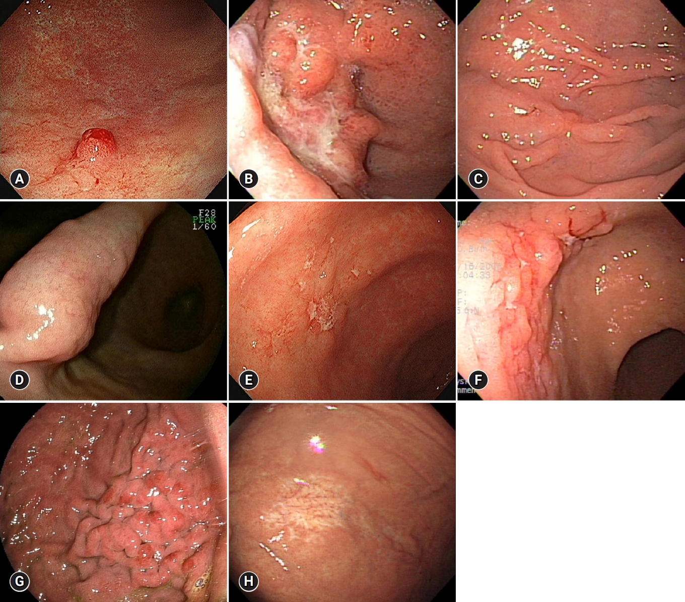
Endoscopic findings of gastric mucosa-associated lymphoid tissue lymphoma. (A) Exophytic type. (B) Ulceroinfiltrative type. (C) IIc-like type. (D) Submucosal tumor type. (E) Multiple erosion type. (F) Cobblestone mucosa type. (G) Partial fold swelling type. (H) Discoloration type.
Endoscopic biopsy using forcep and histopathologic examination are the most basic tests for diagnosis of gastric MALT lymphoma. However, false-negative results in the histologic examination may be possible because the tumor cell of gastric MALT lymphoma originates from the deep mucosa or submucosa and grows without destroying the foveolar gland, which is the basic structure of the mucosal surface [19,20]. Therefore, repeated EGD and endoscopic biopsy may be needed for accurate diagnosis of gastric MALT lymphoma. Furthermore, because gastric MALT lymphoma may have a multifocal distribution of tumors and high-grade gastric MALT lymphoma may be present together, multiple endoscopic biopsies should be performed in the normal mucosa of the antrum, greater and lesser curvatures of the body, and fundus, as well as the abnormal mucosa of at least two tissues in each part [21]. More invasive tissue biopsy such as endoscopic mucosal resection (EMR) or endoscopic submucosal dissection (ESD) may be required if the diagnosis is not confirmed by endoscopic biopsy [22].
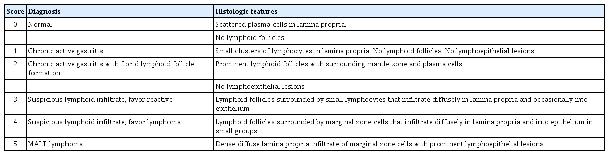
Lymphoepithelial lesion is characteristic feature of gastric MALT lymphoma. Wotherspoon et al. [23] proposed a histological classification system to differentiate normal gastric mucosa and gastric MALT lymphoma (Table 1). Gastric MALT lymphoma can be diagnosed if a histopathologic change corresponding to a score of 5 is observed. If the score is 3 or 4, diagnosis is confirmed based on the monoclonality of B-cells in polymerase chain reaction (PCR) [24]. In addition to morphological evaluation of the tissues, immunohistochemical staining of cluster of differentiation (CD) 20, CD79a, CD43, and more can be performed to diagnose gastric MALT lymphoma. A balanced translocation, t(11;18)(q21;q21), is found in 25–30% of gastric MALT lymphoma cases [25], and it was shown to be associated with a low response to H. pylori eradication therapy and poor prognosis [26,27]. Moreover, t(11;18)(q21;q21) assessed by fluorescence in situ hybridization or API2/MALT1 fusion gene, which is the result of chromosomal translocation, detected by reverse transcriptase PCR, can identify the patients who are expected to have low therapeutic response to H. pylori eradication therapy.
Confirming H. pylori infection in gastric MALT lymphoma is important because the presence of H. pylori affects treatment strategy and response. H. pylori infection must be detected by rapid urease test, histological examination, culture test, urea breath test, and stool antigen test [28], and the presence of H. pylori should be confirmed during the histopathologic evaluation for the diagnosis of gastric MALT lymphoma. Because hematoxylin and eosin staining has low sensitivity for H. pylori diagnosis and has difficulty in detecting non-spiral and spherical H. pylori, special stainings, such as Giemsa, Warthin–Starry, and Alcian blue, or immunostaining for H. pylori, which is the most sensitive and specific, may be required to confirm H. pylori infection [29,30].
Recently, several studies have been reported to improve the diagnosis of gastric MALT lymphoma by using imaging-enhanced endoscopy, such as narrow-band imaging or linked color imaging [31,32].
2. Endoscopic ultrasonography
Endoscopic ultrasonography (EUS), which has sensitivity of 89%, a specificity of 97%, and a total accuracy of 97%, is the most accurate test to assess the depth of tumor invasion of gastric MALT lymphoma [33,34]. EUS is also useful for evaluating local lymph node metastasis. EUS findings of gastric MALT lymphoma can be classified into four types: superficially spreading, diffusely infiltrating, mass forming, and mixed. Superficially spreading and diffusely infiltrating types are seen in low-grade gastric MALT lymphoma, and tumor invasion into the whole gastric wall and lymph node enlargement are characteristics of high-grade gastric MALT lymphoma [35].
EUS can be used to diagnose gastric MALT lymphoma. A thickened gastric wall of 6–12 mm in the EUS can predict gastric MALT lymphoma, and EUS–guided fine needle aspiration (EUS–FNA) can provide diagnostic information by securing false negatives that may occur in endoscopic biopsy [36,37].
EUS can help predict the treatment response of gastric MALT lymphoma. When the gastric MALT lymphoma infiltrates into the submucosal layer in EUS, the complete remission rate for H. pylori eradication therapy is lower than that for the mucosal layer. When the tumor invades the muscle layer, complete remission was not achieved by H. pylori eradication therapy [38,39] (Fig. 2).

Endoscopic ultrasonography findings of gastric mucosa-associated lymphoid tissue lymphoma. (A) Tumor limited to the mucosa. (B) Tumor invading the submucosa.
After the treatment of gastric MALT lymphoma, therapeutic response or relapse after the remission could be evaluated by tracking the changes in the gastric wall thickness by EUS [40]. However, even when remission is achieved, normalization of the thickened gastric wall occurs slowly, and EUS may show normal findings even if the tumor remains histologically [41]. Nevertheless, if thickened stomach wall is observed on EUS even after sufficient time has elapsed since treatment of gastric MALT lymphoma, the possibility of residual gastric MALT lymphoma cannot be ruled out, albeit normal histopathologic examination results [35].
Follow-up of gastric MALT lymphoma
Complete remission of gastric MALT lymphoma can be achieved at 80–86% by H. pylori eradication therapy [42,43]. H. pylori eradication therapy is also considered as a primary treatment option for H. pylori-negative gastric MALT lymphoma [44]. H. pylori eradication was confirmed at least 4 weeks after H. pylori eradication therapy, and follow-up test with EGD, histology, and EUS were performed every 3 months until complete remission was achieved. Complete remission after H. pylori eradication therapy is slow and may take more than a year in some cases [45]. The number of tumor cells is decreased after H. pylori eradication therapy; hence, a sufficient amount of biopsy specimens must be obtained. Complete remission can be determined if histologic remission is identified in two consecutive biopsy results.
After remission, lesions of gastric MALT lymphoma changed to atrophic and whitish discolored mucosa [46] (Fig. 3). Recovery of gastric pit and microvascular structures, which were destroyed and disappeared, can be seen on magnifying endoscopy [47]. The whitish discolored mucosa with definite vascular network showed no recurrence after remission, but the granular mucosa without definite vascular network more likely recurs after remission [48].
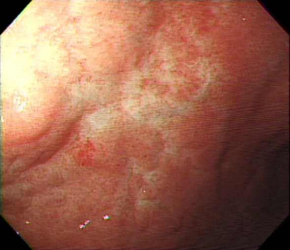
Endoscopic findings of gastric MALT lymphoma after treatment. Mucosa, which was diagnosed as MALT lymphoma, had changed to atrophic and whitish discolored mucosa after Helicobacter pylori eradication. MALT, mucosa-associated lymphoid tissue.
After remission, gastric MALT lymphoma can recur regardless of H. pylori reinfection, and it has a long-term recurrence rate of 7–8%, and the recurrence rate is 2.2% annually [9,42]. Because the risk of developing metachronous gastric adenocarcinoma is increased and this risk persists after several years in patients with gastric MALT lymphoma [49], annual follow-up EGD is recommended after remission of gastric MALT lymphoma. However, there is a lack of studies about how long follow-up EGD should be performed. Because the endoscopic findings of recurred gastric MALT lymphoma are non-specific and variable, sufficient biopsies at multiple sites must be performed.
Conclusion
The early diagnosis of gastric MALT lymphoma is difficult because its symptoms and endoscopic findings are nonspecific. Therefore, if an abnormal mucosa is observed during EGD, gastric MALT lymphoma should be considered, and multiple biopsies and EUS should be performed. In addition, the possibility of false-negative results of endoscopic biopsy should be considered, and more invasive tissue biopsy, such as EUS-FNA, EMR, and ESD, may be necessary. EUS can be used not only for diagnosis, but also for prediction of treatment response. Regular follow-up EGD and EUS are required after treatment of gastric MALT lymphoma.
Acknowledgements
This work was supported by a grant from Chunma Medical Research Foundation, Korea, 2017.
Notes
No potential conflicts of interest relevant to this article was reported.