Breast implant-associated anaplastic large-cell lymphoma (BIA-ALCL)
Article information

Abstract
Breast implant-associated anaplastic large-cell lymphoma (BIA-ALCL) is a rare T-cell non-Hodgkin lymphoma characterized as CD30 positive and anaplastic lymphoma kinase (ALK) negative. In 2016, the World Health Organization declared BIA-ALCL as a new disease entity. The first case of BIA-ALCL was reported in 1997, and as of July 2019, the United States Food and Drug Administration had cited a total of 573 United States and global medical device reports of BIA-ALCL, including 33 deaths. In all clinical case reports, except for those with unknown clinical history, the patient had received at least one textured surface breast implant. Although the etiology is not yet clear, chronic inflammation has been proposed as a potential precursor to tumorigenesis. The most common presentation of BIA-ALCL is peri-implant fluid collection following aesthetic or reconstructive implantation with textured surface breast implants. It can be accompanied by breast swelling, asymmetry, pain, skin lesions, lymphadenopathy, and B-type symptoms. Most cases are detected on average 7 to 10 years after implantation. Diagnostic specimens can be obtained with fine-needle aspiration or biopsy. BIA-ALCL is CD30 positive, epithelial membrane antigen positive, and ALK negative. It can be cured with complete surgical excision at the T1–T3 stage.
Introduction
In August 2019, the Ministry of Food and Drug Safety of Korea (MFDS) ordered to stop using and selling Biocell textured breast implants (Allergan Inc., Irvine, CA, USA). This decision came after the first case of breast implant-associated anaplastic lymphoma (BIA-ALCL) in Korea was reported on August 14, 2019 [1]. As of now, three cases of BIA-ALCL have been reported in South Korea [2]. BIA-ALCL is an uncommon T-cell non-Hodgkin lymphoma characterized as CD30 positive and anaplastic lymphoma kinase (ALK) negative. Primary lymphoma of the breast is very rare, accounting for only 0.12% to 0.53% of all malignant breast tumors, approximately 2.2% of extranodal lymphomas, and less than 1% of all non-Hodgkin lymphomas. Over the past decades, there has been increasing doubt about an etiologic link between breast implants and the development of ALCL. In January 2011, the United States (US) Food and Drug Administration (FDA) stated the relationship between BIA-ALCL and breast implants for the first time as “Although ALCL is extremely rare, the FDA believes that women with breast implants may have a very small but increased risk of developing this disease in the scar capsule adjacent to the implant” [3]. In 2016, the World Health Organization declared BIA-ALCL as a new disease entity [4].
Epidemiology
The first case of BIA-ALCL was reported by Keech and Creech [5] in 1997, and by July 2019, the FDA updated a total of 573 US and global medical device reports of BIA-ALCL, including 33 deaths (Tables 1, 2) [6]. Because of its rare occurrence, it is difficult to determine the exact prevalence of BIA-ALCL. In 2008, de Jong et al. [7] published the first case-control study and reported that the risk of BIA-ALCL development in women with breast implants was 18.2-fold higher than in women who did not have implants (odds ratio, 18.2; 95% confidence interval [CI], 2.1–156.8). In 2018, the same group reported the relative risk of BIA-ALCL with breast implants as 421.8 (95% CI, 526.6–3,385.2) and absolute cumulative risks of 29 per million and 82 per million at 50 years and 70 years, respectively. The estimated prevalence of BIA-ALCL with breast implants in women aged 20 to 70 years was 3.3% [7]. In 2017, Doren et al. [8] published the first US population-based report demonstrating a significant association between textured breast implants and BIA-ALCL. They reported that BIA-ALCL develops only with textured implants, with an incidence rate of 2.03 per million per year, which is 67.6 times higher than that of primary ALCL of the breast in the general population. Lifetime prevalence was estimated to be 33 per million (one per 30,000) women with textured breast implants [8]. In 2017, a study from Australia and New Zealand reported the highest incidence of BIA-ALCL. Fifty-six cases in total had been confirmed by 2017, including 26 new cases of BIA-ALCL diagnosed between January 2017 and April 2018, representing a 47% increase in the number of confirmed cases. The estimated incidence has subsequently been revised from one in 300,000 to one in 1,000–10,000 patients [9]. The number of cases is increasing with the growing interest and recognition among physicians.

Summary of US and global deaths reported in MDRs received as of July 6, 2019 (n=33)

Summary of US and global data as of July 6, 2019 (n=573)
Implant texture and manufacturer
All clinical case reports have demonstrated a strong relationship between BIA-ALCL and textured breast implants. In 2015, Brody et al. [10] reviewed all current BIA-ALCL literature, analyzing 173 cases of the disease, and found that all patients with a known clinical history had received at least one textured surface implant. Additionally, there were no cases before the introduction of textured surface implants. Of the 573 US and global medical device reports of BIA-ALCL, 385 cases had a history of textured implants, 162 were not specified, and 26 had smooth implants. However, these 26 smooth implant cases either had a history of prior exposure to textured implant before revision surgery to smooth implants or no clinical history to review (Table 2) [6].
Magnusson et al. [11] investigated the implant-specific risks of BIA-ALCL with 110 implants in 81 patients in 2019. They reported that the implant-specific risk is 23.4 times higher with Silimed polyurethane (Silimed, Rio De Janeiro, Brazil) and 16.52 times higher with Biocell implants, compared with Siltex implants (Mentor, Santa Barbara, CA, USA). A total of 484 cases of the 573 (84.5%) registered the US and global medical device reports had a history of Allergan implants. Of the 33 reported deaths, no information regarding the implant manufacturer was available for 20 of them; among the remaining 13, 12 had Allergan implants. This is the reason why MFDS ordered a ban on Biocell breast implants (Tables 1, 2). The FDA recalled Allergan textured breast implants and expanders because of the higher rate of BIA-ALCL associated with Biocell breast implants.
Etiology
The etiology and process of BIA-ALCL development are not well understood, but it is likely a complex process involving multiple factors. However, it is related to textured implants, and chronic inflammation has been proposed as a potential etiologic factor and precursor to tumorigenesis. Various pathogenetic theories, including the immunologic hypothesis, tribology, and subclinical infection, have been proposed to explain the mechanism of chronic inflammation. The immunology hypothesis suggests that silicone particles released from the surface of textured implants generate foreign bodies, resulting in chronic inflammation [12]. According to the tribology hypothesis, aggressively textured implants cause delamination of the periprosthetic capsule and lead to the formation of a double capsule through mechanical tear stress [13], consequently causing unresolved inflammation, genetic instability, and activation of maladaptive homeostatic responses and dormant transcription factors [14]. The subclinical infection hypothesis was supported by studies carried out by Hu et al. [15] in 2015. in 2014. He compared the microbiological colonization of implant capsules between BIA-ALCL patients and patients with capsular fibrosis. The BIA-ALCL groups had a higher bacterial burden and a significantly different distribution of bacteria, predominated by the gram-negative pathogen Ralstonia pickettii.
Clinical presentation
The first and most common symptom of BIA-ALCL is unilateral or bilateral peri-implant fluid collection following aesthetic or reconstructive implantation with textured surface breast implants. It can be accompanied by breast swelling, asymmetry, or pain. Skin symptoms (e.g., inflammation, papules) and unilateral regional lymphadenopathy have been described [16]. B-type symptoms such as fever, lymphadenopathy, night sweating, and fatigue can be accompanied [17]. Most cases of BIA-ALCL are detected on average 7 to 10 years after implantation. However, there was one reported within 2 years, and another reported as late as 32 years after implantation [18]. In addition, there was one occurrence 2 months after the exchange of an implant [17]. Most cases are unilateral, but four cases of bilateral involvement have been reported in patients with bilateral breast implants [19]. Approximately 60% of patients present with malignant effusion, 17% with a mass, and 20% of patients present with both seroma and mass [20].
Diagnosis
Standardized diagnosis and management guidelines for BIA-ALCL have been established by the National Comprehensive Cancer Network (NCCN) [21]. BIA-ALCL should be suspected and evaluated for patients who develop spontaneous peri-implant fluid collection occurring more than 1 year after aesthetic or reconstructive implantation with a textured surface breast implant. Many patients with breast implants are likely to have a small amount of peri-implant fluid (5–10 mL) without symptoms. These are normal findings and do not require further evaluation. Ultrasonography is the best imaging method for detecting and defining any peri-implant fluid or mass. Suspicious fluid collections should be aspirated with a fine needle under ultrasonography guidance. A minimum of 10 mL (ideally >50 mL) of fluid should be collected to diagnose BIA-ALCL. A suspected mass requires a tissue biopsy. Specimens should be sent for cell morphology by cytology, immunohistochemistry, and flow cytometry [22-25]. BIA-ALCL is CD30 positive, epithelial membrane antigen positive, and ALK negative (Fig. 1). After histologic confirmation of BIA-ALCL, further lymphoma workup and staging are recommended. Each case should ideally be discussed at a multidisciplinary team conference consisting of oncologists, radiologists, pathologists, and plastic surgeons. Routine laboratory work should include complete blood cell count with differential, comprehensive metabolic panel, and lactate dehydrogenase levels. Positron emission tomographic (PET) and computed tomographic (CT) scans are beneficial for demonstrating associated capsular masses, chest wall involvement, regional lymphadenopathy, and/or distant organ metastasis [10].
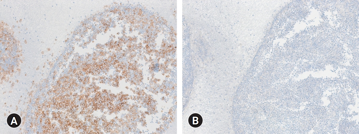
(A) Immunohistochemistry for CD30 highlights the membranes of neoplastic cells. (B) The neoplastic cells are negative for anaplastic lymphoma kinase (ALK) (immunohistochemical stain, x100 [A] and [B]).
Preoperative evaluation/staging
There are two main staging systems for BIA-ALCL, the Lugano modification of the Ann Arbor staging system and the BIA-ALCL tumor, node, and metastasis (TNM) staging system. The traditional staging system for non-Hodgkin lymphoma is the Lugano modification of the Ann Arbor staging system, which has been used in many previous reports. In this system, stage IE disease is limited to a single extranodal (E) site such as the breast or implant capsule, whereas stage IIE disease is defined as an extranodal disease with spread to or involvement of local lymph nodes [26]. Most patients with BIA-ALCL have an early-stage disease, either stage IE (83%–84%) or stage IIE (10%–16%), while a few of them (0%–7%) fall into stage IV disease with this system [21,27,28]. Because of the unique characteristics of BIA-ALCL that behaves like a solid tumor rather than a liquid tumor and that the Ann Arbor staging system does not consider capsular invasion, NCCN is now using the TNM staging system modeled after the American Joint Committee on Cancer TNM [27]. However, the TNM classification describes BIA-ALCL as a spectrum of disease from stage IA (35%–70%, effusion only), IB (3%–11%), IC (8%–13%), IIA (8%–25%) [7,14,21], IIB (3%–5%), and III (3%–9%) to IV (1%–2%) [7,27].
Treatment
The most important factors for the treatment of BIA-ALCL are timely diagnosis and complete surgical excision [27]. The goals of surgery are complete removal of the implant, including the surrounding fibrous capsule and any associated mass. Complete surgical excision prolongs overall survival and event-free survival compared with all other therapeutic interventions. In subpectoral implant placement, adherence to the rib cage can make complete resection difficult, while an injection of tumescent solution facilitates complete excision. In this case, care must be taken to avoid pneumothorax. The effect of local seeding of malignant seroma on capsulectomy is not yet clear. However, clinically, this has not been observed to influence the recurrence rate. In cases presenting with a mass, complete excision of the mass with a negative margin is essential. At present, there is no clear role for radical mastectomy or sentinel lymph node biopsy. Full axillary dissection has rarely been used for the gross involvement of multiple lymph nodes. According to the NCCN guidelines, an estimated 2% to 4% of patients develop bilateral disease, and therefore surgeons may consider the removal of the contralateral implant. The rate of disease events and recurrence is 2.6-fold higher for stage II disease and 2.7-fold higher for stage III disease than for stage I disease [26]. The recurrence rate following complete surgical excision is 14.3% for patients with T4 disease compared with 0% for patients with T1 to T3 disease [27].
Adjuvant therapy
There are no established treatment protocols for stage II or more advanced, disseminated, and recurrent cases after complete resection. Therefore, treatment protocols for primary cutaneous and systemic ALCL are generally used. Radiation therapy with 24 to 36 Gy is suggested for patients with local residual disease, positive margins, or unresectable disease with chest wall invasion like the cutaneous ALCL [29]. Systemic therapy combined with anthracycline-based chemotherapy or brentuximab vedotin is used for stage II or more advanced or disseminated state [30-35].
Follow-up
Patients showing complete response to treatment can be monitored with history and physical exanimation every 3 to 6 months for 2 years and then as clinically indicated. Monitoring may include CT or PET/CT scans every 6 months for 2 years and then only if clinically indicated.
Conclusion
As of 2020, three cases of BIA-ALCL have been reported in Korea. This means that Korea is no longer a safe country from BIA-ALCL, and more patients may be reported. Every symptomatic peri-implant fluid collection for more than 1 year after textured surface implantation with aesthetic or reconstructive surgery should be evaluated for BIA-ALCL.
Notes
Conflicts of interest
No potential conflict of interest relevant to this article was reported.
Acknowledgements
I thank Professor Il-Kug Kim, Department of Plastic and Reconstructive Surgery, Yeungnam University College of Medicine, for providing the photographs.