PubMed Central, CAS, DOAJ, KCI

Articles
- Page Path
- HOME > J Yeungnam Med Sci > Volume 39(2); 2022 > Article
-
Case report
Palisaded encapsulated neuroma on the lower lip: a case report -
Jung Eun Seol1,2
 , Seong Min Hong1
 , Sang Woo Ahn1
 , Jong Uk Kim1
 , Woo Jung Jin1
 , So Hee Park1
 , Hyojin Kim1
-
Journal of Yeungnam Medical Science 2022;39(2):168-171.
DOI: https://doi.org/10.12701/yujm.2021.01088
Published online: July 9, 2021
1Department of Dermatology, Inje University Busan Paik Hospital, Inje University College of Medicine, Busan, Korea
2Cardiovascular and Metabolic Disease Center, Inje University, Busan, Korea
- Corresponding author: Hyojin Kim, MD, PhD Department of Dermatology, Inje University Busan Paik Hospital, Inje University College of Medicine, 75 Bokji-ro, Busanjin-gu, Busan 47392, Korea Tel: +82-51-890-6135 Fax: +82-51-890-5845 E-mail: derma09@hanmail.net
Copyright © 2022 Yeungnam University College of Medicine, Yeungnam University Institute of Medical Science
This is an Open Access article distributed under the terms of the Creative Commons Attribution Non-Commercial License (http://creativecommons.org/licenses/by-nc/4.0/) which permits unrestricted non-commercial use, distribution, and reproduction in any medium, provided the original work is properly cited.
- 5,429 Views
- 81 Download
Abstract
- Palisading encapsulated neuroma is a rare, benign, cutaneous nerve sheath tumor. It usually occurs as an asymptomatic solitary skin-colored papule and commonly affects the nose and cheeks. Sometimes, it involves other sites, including the shoulder, upper arm, and trunk, but rarely involves the oral mucosa, including that of the lip. In our case, a 63-year-old female patient complained of a pinkish rubbery nodule on her lower lip. Histopathologic examination demonstrated a well-circumscribed nodule encapsulated by connective tissue stroma in the dermis. The nodule consisted of palisading spindle-shaped tumor cells with wavy and basophilic nuclei. The cells were arranged in streaming fascicles with multiple clefts and were strongly positive for S-100 proteins. To our knowledge, only three cases of palisading encapsulated neuroma on the lower lip have been reported in the Korean literature. Herein, we report a rare case of an oral palisaded encapsulated neuroma.
- Palisaded encapsulated neuroma (PEN) or solitary circumscribed neuroma is a benign neural tumor first described by Reed et al. [1] in 1972. It mainly occurs as a single, asymptomatic papule or nodule that slowly grows on the face of a middle-aged adult, and rarely occurs in the oral cavity, shoulder, trunk, limbs, etc. [2]. Histopathologic examination reveals an encapsulated dermal nodule comprised of proliferating Schwann cells and axons arranged in bundles. A total of three cases of PEN on the lips have been reported in South Korean literature [3-5]. It is assumed that this is due to the characteristic of PEN, which is asymptomatic and does not occur in the oral cavity. Herein, we report a rare case of PEN on the lips, along with a review of clinical and histological features.
Introduction
- Ethical statesments: This study was approved by the Institutional Review Board (IRB) of Inje University Busan Paik Hospital (IRB No: BPH 2021-01-001). Written informed consent was obtained from the patient for the publication of this case report and accompanying images.
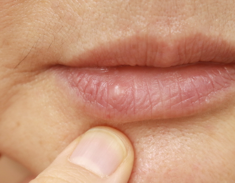
- A 63-year-old female patient presented with a cystic nodule on her lower lip. The lesion persisted for 6 months and gradually increased in size. The patient had no symptoms. She had a history of hypertension, for which she was taking oral medications. There was no significant family history or history of trauma to the lip. Physical examination revealed a solid and rubbery 0.4×0.4 cm nodule on her lower lip (Fig. 1). The nodule was completely excised, and biopsy was performed. Histological examination revealed a nodular tumor in the dermis. The tumor was well-circumscribed and partially encapsulated by fibrous connective tissue (Fig. 2A). Spindle-shaped cells were clustered inside the tumor, and the fascicles of spindle cells were separated by prominent clefts. There was no Verocay body formation, pleomorphism, or mitotic activity (Fig. 2B). On immunohistochemical examination, except for the connective tissue of the capsule, the tumor cells showed a positive reaction to S-100 proteins (Fig. 2C, 2D) and focally positive to neurofilament proteins (Fig. 2E). The above clinical and histological findings led to the diagnosis of PEN. No recurrence was observed after the complete excision.
Case
- PEN is a benign neurogenic tumor that was first described by Reed et al. [1] in 1972. They categorized benign neurogenic tumors into three types; schwannomas, neurofibromas, and neuromas. PEN is a type of neuroma characterized by spindle-shaped cells arranged in a palisading pattern and surrounded by a connective tissue capsule [1]. Clinically, PEN appears as pinkish papules or nodules in middle-aged adults, with no sex predilection [6]. It originates from the peripheral nerves of the skin; therefore, it can occur in all areas where these nerves are distributed. However, in most cases, it occurs on the face over the cheeks and nose. It rarely occurs on the trunk, penis, extremities, and oral cavity, including the lips [7-9]. PEN accounts for approximately 25% of the nerve sheath tumors in the skin. However, it accounts for 11% of nerve sheath tumors in the oral cavity, of which 10% to 16% occur on the lips [2,10]. PEN usually does not cause symptoms such as itching or pain and enlarges over several years.
- Pathologically, it is characterized by round or oval nodules with a capsule composed of thin collagen fibers. In the nodule, spindle-shaped cells form bundles with irregular rows. Immunohistochemical examination shows that the capsule is partially positive for epithelial membrane antigen, suggesting that it is derived from peripheral nerves, and the cells in the nodule are positive for S-100 proteins [11]. In our case, the results of immunohistochemical staining were consistent with those of PEN. Some cases of similar lesions with unclear palisading alignment or capsule formation have been reported, and researchers have proposed calling them “solitary circumscribed neuroma” instead of PEN [12]. In the 2013 and 2020 World Health Organization (WHO) classification of tumors of soft tissue and bone, solitary circumscribed neuroma is described as a formal name, and PEN is described as a synonym. However, even after the WHO classification was published, these terms have been used interchangeably [13,14].
- The pathogenesis of PEN remains controversial. Reed et al. [1] suggested that PEN is a part of multiple endocrine neoplasia syndrome type 2. However, further research confirmed that the similarity in the clinical characteristics of PEN and the neuroma in multiple endocrine neoplasia syndrome type 2 was a coincidence in the case described by Reed et al. [1], and that the clinical characteristics of the two neuromas in other cases have shown many differences [2]. Recently, PEN has been accepted as a regenerative tumor secondary to trauma [7,15]. The morphological and cellular distributions of PEN and traumatic neuroma are found to be similar. Leblebici et al. [9] reported that PEN shows an increase in small-diameter peripheral nerve fibers similar to that in traumatic neuroma around the lesion. The lip is frequently damaged by teeth or food unconsciously; hence, we thought that this minor trauma might have led to the occurrence of PEN in our case.
- Clinicopathologically, PEN in the oral cavity can be differentiated from neurofibroma, traumatic neuroma, and Schwann cell tumors. Unlike PEN, a neurofibroma does not have a capsule, the tumor cells are scattered, and a palisading pattern is not observed [13]. A traumatic neuroma appears after a history of trauma and is commonly accompanied by pain, burning sensation, or paresthesia. Additionally, infiltration of inflammatory cells around or inside the tumor cells can help differentiate a traumatic neuroma from PEN [16,17]. Although a Schwann cell tumor is surrounded by a capsule similar to that in PEN, it does not contain axons in the intracapsular part; therefore, it shows negative immunohistochemical staining for neural filaments, and the Antoni type A and B tissue is an important differentiation point.
- PEN is usually treated by simple excision and rarely grows again, even after partial excision. Initially, our provisional diagnosis was a mucocele, which is a common asymptomatic lesion of the lip having a color similar to that of the mucosa. Although oral neural neoplasms are uncommon, they should be included in the differential diagnosis, and an accurate diagnosis should be made after an immunohistochemical examination.
Discussion
-
Conflicts of interest
No potential conflict of interest relevant to this article was reported.
-
Funding
None.
-
Author contributions
Conceptualization; HK, SHP; Supervision HK; Investigation; JES, SMH; Formal analysis: SWA, JUK, WJJ; Writing-original draft: JES, SMH; Writing-review & editing: SWA, JUK, WJJ, SHP, HK.
Notes

- 1. Reed RJ, Fine RM, Meltzer HD. Palisaded, encapsulated neuromas of the skin. Arch Dermatol 1972;106:865–70.ArticlePubMed
- 2. Koutlas IG, Scheithauer BW. Palisaded encapsulated (“solitary circumscribed”) neuroma of the oral cavity: a review of 55 cases. Head Neck Pathol 2010;4:15–26.ArticlePubMedPMC
- 3. Cho YH, Lee JH, Bang DS, Kim YC. A case of palisaded encapsulated neuroma of the lower lip. Korean J Dermatol 2002;40:1552–6.Article
- 4. Cho DY, Kim DH, Choi KC, Chung BS. A case of palisaded encapsulated neuroma of the lower lip. Ann Dermatol 2006;18:37–9.Article
- 5. Lee SW, Yi JY, Cho BK, Houh W, Kim TY, Song KY. Palisaded encapsulated neuroma. Ann Dermatol 1990;2:136–40.Article
- 6. Manchanda AS, Narang RS, Puri G. Palisaded encapsulated neuroma. J Orofac Sci 2015;7:136–9.Article
- 7. Dover JS, From L, Lewis A. Palisaded encapsulated neuromas: a clinicopathologic study. Arch Dermatol 1989;125:386–9.ArticlePubMed
- 8. Val-Bernal JF, Alvarez-Cañas C, Vega A. Palisaded encapsulated neuroma of the glans penis. Urology 1995;45:689–91.ArticlePubMed
- 9. Leblebici C, Savli TC, Yeni B, Cin M, Aksu AE. Palisaded encapsulated (solitary circumscribed) neuroma: a review of 30 cases. Int J Surg Pathol 2019;27:506–14.ArticlePubMed
- 10. Dakin MC, Leppard B, Theaker JM. The palisaded, encapsulated neuroma (solitary circumscribed neuroma). Histopathology 1992;20:405–10.ArticlePubMed
- 11. Megahed M. Palisaded encapsulated neuroma (solitary circumscribed neuroma): a clinicopathologic and immunohistochemical study. Am J Dermatopathol 1994;16:120–5.ArticlePubMed
- 12. Fletcher CD. Solitary circumscribed neuroma of the skin (so-called palisaded, encapsulated neuroma): a clinicopathologic and immunohistochemical study. Am J Surg Pathol 1989;13:574–80.ArticlePubMed
- 13. Scolyer RA, Ferguson PM. Solitary circumscribed neuroma. In: The WHO Classification of Tumours Editorial Board, editors. WHO classification of tumours. Soft tissue and bone tumours. 5th ed. Lyon: IARC Press; 2020. p. 245–6.
- 14. Kutzner H. Solitary circumscribed neuroma. In: Fletcher CD, Bridge JA, Hogendoorn PCW, Mertens F, editors. WHO classification of tumours of soft tissue and bone. 4th ed. Lyon: IARC Press; 2013. p. 181–2.
- 15. Argenyi ZB, Santa Cruz D, Bromley C. Comparative light-microscopic and immunohistochemical study of traumatic and palisaded encapsulated neuromas of the skin. Am J Dermatopathol 1992;14:504–10.ArticlePubMed
- 16. Lopes MA, Vargas PA, Almeida OP, Takahama A, León JE. Oral traumatic neuroma with mature ganglion cells: a case report and review of the literature. J Oral Maxillofac Pathol 2009;13:67–9.ArticlePubMedPMC
- 17. Maharudrappa B, Kumar GS. Neural tumours of oral and para oral region. Int J Dent Clin 2011;3:34–43.
References
Figure & Data
References
Citations


 E-Submission
 Yeungnam University College of Medicine
 PubReader
 ePub Link
 Cite
Cite


