Skip Navigation
Skip to contents

JYMS : Journal of Yeungnam Medical Science

Indexed in: ESCI, Scopus, PubMed,
PubMed Central, CAS, DOAJ, KCI
FREE article processing charge
OPEN ACCESS
SEARCH
Search

Articles

Page Path
HOME > J Yeungnam Med Sci > Volume 40(3); 2023 > Article
Image vignette
Effective treatment of empyema thoracis caused by a ruptured large hepatic cyst
Seok Soo Lee1orcid, Hyuckgoo Kim2orcid
Journal of Yeungnam Medical Science 2023;40(3):315-316.
DOI: https://doi.org/10.12701/jyms.2023.00262
Published online: May 12, 2023

1Department of Thoracic and Cardiovascular Surgery, Yeungnam University College of Medicine, Daegu, Korea

2Department of Anesthesiology and Pain Medicine, Yeungnam University College of Medicine, Daegu, Korea

Corresponding author: Seok Soo Lee, MD Department of Thoracic and Cardiovascular Surgery, Yeungnam University College of Medicine, 170 Hyeonchung-ro, Nam-gu, Daegu 42415, Korea Tel: +82-53-620-3882 Fax: +82-53-620-3880 • E-mail: andrea0710@naver.com
• Received: March 15, 2023   • Revised: April 3, 2023   • Accepted: April 7, 2023

Copyright © 2023 Yeungnam University College of Medicine, Yeungnam University Institute of Medical Science

This is an Open Access article distributed under the terms of the Creative Commons Attribution Non-Commercial License (http://creativecommons.org/licenses/by-nc/4.0/) which permits unrestricted non-commercial use, distribution, and reproduction in any medium, provided the original work is properly cited.

prev
  • 843 Views
  • 34 Download
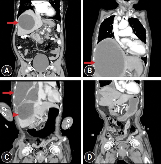
An 87-year-old woman presented to the emergency department with right-sided chest pain and dyspnea. The patient had been undergoing outpatient internal medical follow-up for a hepatic cyst 10 years previously (Fig. 1A). One month before visiting the hospital, she underwent computed tomography (CT) due to pain in the upper abdomen, which revealed that the size of the hepatic cyst had increased considerably (Fig. 1B). However, her symptoms were not severe and the patient was older. She only wanted to control her symptoms. At the time of admission to the emergency room, a large amount of pleural fluid was observed on CT, and the size of the hepatic cyst had decreased (Fig. 1C), resulting in empyema thoracis as the hepatic cyst had ruptured through the diaphragm and into the thoracic cavity [1,2]. The patient was immediately treated with closed-tube thoracostomy. After the procedure, approximately 2,000 mL of pleural effusion was drained, and her dyspnea improved. The pH of the drained pleural fluid was 6.57, with a glucose level of 6 mg/L, lactate dehydrogenase level of 51,361 IU/L, and white cell count of 176,000/μL. Empyema was diagnosed based on these results, and intravenous piperacillin/tazobactam antibiotics were initiated. Although a diaphragmatic defect or fistula was not clearly visible on the CT scan, >150.0 μmol/L of bile acid was found in the pleural fluid, confirming empyema from the hepatic lesion. Five days after the procedure, the patient’s symptoms significantly improved, and a follow-up CT scan was performed. The large hepatic cyst had almost disappeared, and the empyema was well drained (Fig. 1D). Escherichia coli was identified in the pleural effusion, and piperacillin/tazobactam was continued because E. coli is highly sensitive to this antibiotic combination. The chest tube was removed on the 19th day of hospitalization. She was discharged without any discomfort 2 days after removal of the chest tube. On the 9th day after discharge, she visited the outpatient clinic, and chest radiography confirmed that her right lung had improved. It has been 2 years since the patient was treated and discharged from our hospital. She is currently being followed up at the neurology department of a local medical center for dementia. She has had no subsequent symptoms such as dyspnea or abdominal pain.
Hepatic cysts are usually asymptomatic; however, in rare cases, a liver abscess may occur as a complication of an infection. Pyogenic liver abscess (PLA) is primarily caused by bacteria, such as E. coli, Streptococcus spp., and Klebsiella pneumoniae [3]. PLA is rare; however, empyema caused by a liver abscess that bursts through the diaphragm into the chest cavity is much rarer [4]. As the risk of death is high if treatment is not initiated quickly, effective drainage and appropriate use of antibiotics are required to treat patients [3].

Ethical statements

This study was approved by the Institutional Review Board (IRB) of Yeungnam University Hospital (IRB No: YUMC 2023-03-030). Informed consent was waived because of the retrospective nature of the image vignette.

Conflicts of interest

Hyuckgoo Kim has been an editorial board member of Journal of Yeungnam Medical Science since 2021. He was not involved in the review process of this manuscript. There is no conflict of interest to declare.

Funding

None.

Author contributions

Conceptualization, Data curation, Resources: SSL, HK; Formal analysis, Visualization, Software, Supervision: SSL; Writing-original draft: SSL, HK; Writing-review & editing: SSL.

Fig. 1.
(A) Hepatic cyst (10 cm, arrow) noted by abdominal computed tomography (CT) 10 years ago. (B) CT scan taken at the internal medicine outpatient department 1 month before the visit to the emergency room shows the existing hepatic cyst enlarged to approximately 19 cm (arrow). (C) Abdominal CT at the time of admission to the emergency room shows a large amount of pleural effusion (arrow) and the hepatic cyst (arrowhead), which is significantly smaller than 1 month earlier. (D) One week after chest tube insertion, CT scan shows that most of the pleural effusion has drained, and the giant hepatic cyst has disappeared.
jyms-2023-00262f1.jpg
  • 1. Peterson M, Aboudara M. Hepatic cystopleural fistula. Am J Respir Crit Care Med 2014;189:e4–6.ArticlePubMed
  • 2. Ikeda M, Hatakeyama Y, Murakami S, Hashimoto R, Tauchi S, Yonekura Y, et al. Surgical repair of hepatic hydrothorax caused by diaphragmatic fistula. Respir Med Case Rep 2020;32:101325.ArticlePubMedPMC
  • 3. AlGhamdi ZM, Boumarah DN, Alshammary S, Elbawab H. Pleural empyema as a complication of pyogenic liver abscess: can the minimum achieve the optimal? A comparison of 3 approaches. Am J Case Rep 2021;22:e935169.ArticlePubMedPMC
  • 4. Kim DH. Empyema caused by transdiaphragmatic extension of pyogenic liver abscess. Clin Case Rep 2018;7:240–1.ArticlePubMedPMCPDF

Figure & Data

References

    Citations

    Citations to this article as recorded by  

      Figure
      • 0
      Effective treatment of empyema thoracis caused by a ruptured large hepatic cyst
      Image
      Fig. 1. (A) Hepatic cyst (10 cm, arrow) noted by abdominal computed tomography (CT) 10 years ago. (B) CT scan taken at the internal medicine outpatient department 1 month before the visit to the emergency room shows the existing hepatic cyst enlarged to approximately 19 cm (arrow). (C) Abdominal CT at the time of admission to the emergency room shows a large amount of pleural effusion (arrow) and the hepatic cyst (arrowhead), which is significantly smaller than 1 month earlier. (D) One week after chest tube insertion, CT scan shows that most of the pleural effusion has drained, and the giant hepatic cyst has disappeared.
      Effective treatment of empyema thoracis caused by a ruptured large hepatic cyst

      JYMS : Journal of Yeungnam Medical Science
      TOP